Bacteria are a large group of minute, unicellular, microscopic organisms, which have been classified as prokaryotic cells, as they lack a true nucleus. These microscopic organisms comprise a simple physical structure, including cell wall, capsule, DNA, pili, flagellum, cytoplasm and ribosomes.
Bacteria can be gram-positive or gram-negative depending upon the staining methods. Let us have a detailed look at the difference between the two types of bacteria.
Difference between Gram-Positive and Gram-Negative Bacteria
Following are the important differences between gram-positive and gram-negative bacteria:

Difference between Gram-Positive and Gram-Negative Bacteria
| Gram-Positive bacteria | Gram-Negative bacteria |
| Cell Wall | |
| A single-layered, smooth cell wall | A double-layered, wavy cell-wall |
| Cell Wall thickness | |
| The thickness of the cell wall is 20 to 80 nanometres | The thickness of the cell wall is 8 to 10 nanometres |
| Peptidoglycan Layer | |
| It is a thick layer/ also can be multilayered | It is a thin layer/ often single-layered. |
| Teichoic acids | |
| Presence of teichoic acids | Absence of teichoic acids |
| Outer membrane | |
| The outer membrane is absent | The outer membrane is present (mostly) |
| Porins | |
| Absent | Occurs in Outer Membrane |
| Mesosome | |
| It is more prominent. | It is less prominent. |
| Morphology | |
| Cocci or spore-forming rods | Non-spore forming rods. |
| Flagella Structure | |
| Two rings in basal body | Four rings in basal body |
| Lipid content | |
| Very low | 20 to 30% |
| Lipopolysaccharide | |
| Absent | Present |
| Toxin Produced | |
| Exotoxins | Endotoxins or Exotoxins |
| Resistance to Antibiotic | |
| More susceptible | More resistant |
| Examples | |
| Staphylococcus, Streptococcus, etc. | Escherichia, Salmonella, etc. |
| Gram Staining | |
| These bacteria retain the crystal violet colour even after they are washed with acetone or alcohol and appear as purple-coloured when examined under the microscope after gram staining. | These bacteria do not retain the stain colour even after they are washed with acetone or alcohol and appear as pink-coloured when examined under the microscope after gram staining. |
Gram-Positive and Gram-Negative Bacteria – Overview
The gram-positive bacteria retain the crystal violet colour and stains purple whereas the gram-negative bacteria lose crystal violet and stain red. Thus, the two types of bacteria are distinguished by gram staining.
Gram-negative bacteria are more resistant against antibodies because their cell wall is impenetrable.
Gram-positive and gram-negative bacteria are classified based on their ability to hold the gram stain. The gram-negative bacteria are stained by a counterstain such as safranin, and they are de-stained because of the alcohol wash. Hence under a microscope, they are noticeably pink in colour. Gram-positive bacteria, on the other hand, retains the gram stain and show a visible violet colour upon the application of mordant (iodine) and ethanol (alcohol).
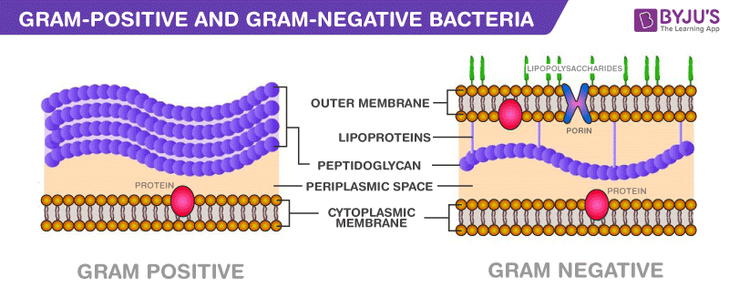
Gram-positive bacteria constitute a cell wall, which is mainly composed of multiple layers of peptidoglycan that forms a rigid and thick structure. Its cell wall additionally has teichoic acids and phosphate. The teichoic acids present in the gram-positive bacteria are of two types – the lipoteichoic acid and the teichoic wall acid. The cell wall is known as murein.
In gram-negative bacteria, the cell wall is made up of an outer membrane and several layers of peptidoglycan. The outer membrane is composed of lipoproteins, phospholipids, and LPS. The peptidoglycan stays intact to lipoproteins of the outer membrane that is located in the fluid-like periplasm between the plasma membrane and the outer membrane. The periplasm is contained with proteins and degrading enzymes which assist in transporting molecules.
The cell walls of the gram-negative bacteria, unlike the gram-positive, lacks the teichoic acid. Due to the presence of porins, the outer membrane is permeable to nutrition, water, food, iron, etc.
Gram Staining
This technique was proposed by Christian Gram to distinguish the two types of bacteria based on the difference in their cell wall structures. The gram-positive bacteria retain the crystal violet dye, which is because of their thick layer of peptidoglycan in the cell wall.
This process distinguishes bacteria by identifying peptidoglycan that is found in the cell wall of the gram-positive bacteria. A very small layer of peptidoglycan is dissolved in gram-negative bacteria when alcohol is added.
Difference between Gram-Positive and Gram-Negative Bacteria – Key Points
- The cell wall of gram-positive bacteria is composed of thick layers peptidoglycan.
- The cell wall of gram-negative bacteria is composed of thin layers of peptidoglycan.
- In the gram staining procedure, gram-positive cells retain the purple coloured stain.
- In the gram staining procedure, gram-negative cells do not retain the purple coloured stain.
- Gram-positive bacteria produce exotoxins.
- Gram-negative bacteria produce endotoxins.
For more information on the differences between gram-positive and gram-negative bacteria, keep visiting BYJU’S website or download the BYJU’S app for further reference.
Further Reading:
Frequently Asked Questions
Give a few examples of gram-positive bacteria.
Gram-positive bacteria include the bacteria of genre Staphylococcus, Streptococcus, Enterococcus. These bacteria are the most common cause of clinical infections.
Which is more harmful- gram-positive bacteria or gram-negative bacteria?
Gram-negative bacteria are more harmful and cause certain diseases. Their outer membranes are hidden by a slime layer that hides the antigens present in the cell.
Is it easier to kill gram-positive bacteria?
The cell wall of the gram-positive bacteria absorbs antibiotics and cleaning products. Because of the outer peptidoglycan layer, they are easier to kill. Gram-negative bacteria cannot be killed easily.
What infections are caused by gram-positive bacteria?
Gram-positive bacteria usually cause Urinary Tract Infections. These are caused commonly in people who are more prone to urinary tract infections or are elderly or pregnant.
Which infections are caused by gram-negative bacteria?
The gram-negative bacteria cause various infections in humans such as indigestion, food poisoning, pneumonia, meningitis and other bacterial infections in the blood cells, bloodstream, wound infections, etc. The infections are caused by Acinetobacter, Pseudomonas aeruginosa and E.coli.


It’s a good understanding distinguish between gram +ve & -ve bacteria . also helpful to us.
I̮t̮’s̮ a̮ v̮e̮r̮y̮ e̮a̮s̮y̮ l̮a̮n̮g̮u̮a̮g̮e̮ t̮o̮ u̮n̮d̮e̮r̮s̮t̮a̮n̮d̮ s̮o̮m̮e̮ i̮m̮p̮o̮r̮t̮a̮n̮t̮ d̮i̮f̮f̮e̮r̮e̮n̮c̮e̮s̮ b̮e̮t̮w̮e̮e̮n̮ g̮r̮a̮m̮ +v̮e̮ & g̮r̮a̮m̮ -v̮e̮ b̮a̮c̮t̮e̮r̮i̮a̮
THANKS ALOT I GET GOOD UNDER STANDING
it’s very helpful contents thank you for these value informations
Very helpful indeed!
Thanks for the information
Information is very helpful in plant pathology studies.
It is clear information about gram positive vs gram negative bacteria. Thanks a lot!
Helpful